Translocation between chromosomes 1 and 8 in cattle, 2n=60,XY. Staining method: BrdU incorporation - Acridine Orange. .

If an individual does not have a balanced set of chromosomes, i.e. two homologue chromosomes in each pair (and for the male an X and a Y), this will normally be visible in the phenotype, which then shows more or less deviation from normality. Animals with a non-balanced set of chromosomes will most often be sterile and have low vitality. Animals with a balanced set of chromosomes will generally be normal phenotypically. Chromosome deviations, in animals with a normal phenotype, are normally detected due to low fertility or complete sterility. The karyotype of a bull with low fertility having a 1/8 translocation is shown in Figure 10.2, cf. Christensen, K., Agerholm, J.S. & Larsen, B. 1992a. Dairy breed bull with complex chromosome translocation: Fertility and linkage studies. Hereditas 117, 199-202.
| Figure 10.2 Translocation between chromosomes 1 and 8 in cattle, 2n=60,XY. Staining method: BrdU incorporation - Acridine Orange. . |
 |
Other types of chromosome deviations worth mentioning are trisomies, the best
known case being trisomy 21 in human. It occurs in about one in 500 new born
babies.
The trisomies are very rare in animals, but they occasionally occur.
Below is shown a trisomy 28 in cattle. The animal suffered from cleft
palate and heart abnormalities, see
Figure 10.3.
| Figure 10.3. Trisomy 28 in a calf, live born but unable to survive,
2n=61,XX. Staining method: BrdU incorporation - Acridine Orange. |
 |
Normally the foetuses carrying trisomy 28 are aborted or die straight after birth.
In most domestic animals less severe chromosome errors occur. For instance fusion of two acrocentric
chromosomes, called centric fusion.
The best known is the 1/29 centromere fusion of chromosome 1 and 29 in cattle. When such a fusion occurs in
heterozygote form, it causes a slightly lower fertility of about 10 %, when measured as
return to the service rate.
A corresponding centromere fusion is found in the blue foxes. Here the effect is of the same magnitude concerning litter size, see table below where the chromosome number 49 corresponds to heterozygotes with respect to the centromere fusion. Data from Christensen et al. Hereditas 1982, 97:211-215.
Dam chromosome number  Number of litters    Litter size
      48                  16                    11.9
      49                  42                     9.8
      50                  17                    11.2

Sire chromosome number
      48                  10                    12.2
      49                  52                    10.1
      50                  13                    11.2
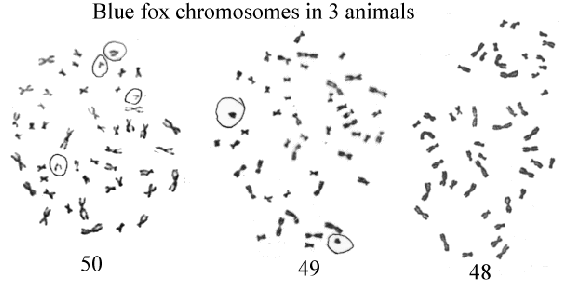
Metaphase chromosomes of the three types are shown in Figure 10.4. In the figure there is a circle around the chromosomes that participate in the centromere fusion.
| Figure 10.4. A centromere fusion of two chromosomes results in varying chromosome numbers between 48 and 50 in the blue fox |
 |
It is known that some mice carry up to seven sets of centromere fusions. When all fusions occur in heterozygote form these mice have a very low fertility, consistent with a reduction in fertility of about 10 % per centromere fusion in the heterozygote stage.
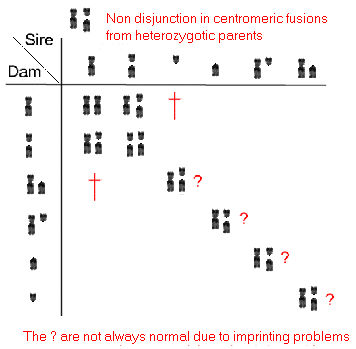
Figur 10.4a. Segregations of chromosomes from centromere fusion heterozygotes result in non disjunction and uniparantal disomy.  |
Free martins: Chromosome investigations can be used to identify animals with placental anastomoses, which often occur in cattle twins. When mixing the blood in the early foetal stages, a mixture of stem cells are established for the white and the red blood cells. The proportions are from 0 to 100 % of the right 'type'. If the mixing is too extensive the heifer in a mixed twin pair gets abnormal sexual organs and is infertile. The bull calf has normal fertility, but in paternity testing by means of a blood sample mistakes can occur, as this might show the genotype of the other twin. Therefore, when delivering blood samples for a paternity tests information should be given, if one of the involved animals have a twin.
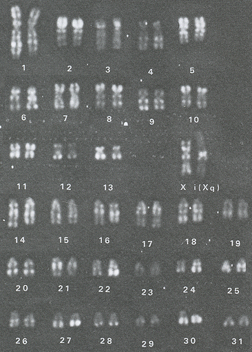
| Figure 10.5. Horse chromosomes 64, XX. One of the X-chromosomes has two
q-arms. It was always inactive. |
 |
Sex-chromosome errors in mares: Mares with XY sex-chromosomes are fairly common in some of the half-breeds. They are not fertile. There also occur a fair number of mares with XO or XXX sex-chromosome constellations. Figure 10.5 shows chromosomes of a mare with an abnormal X chromosome, which is always inactive. It has two q-arms. The picture is provided by I. Gustavsson, The Swedish Agricultural University.
Auli Mäkinä, at the university of Helsinki has a good survey over chromosome aberrations in the horse.
Sister chromatid exchange (SCE)
Cells which are grown for two cycles, BrdU being added in the first cycle, can reveal SCE. SCE can identify individuals with unstable chromosomes or be utilized
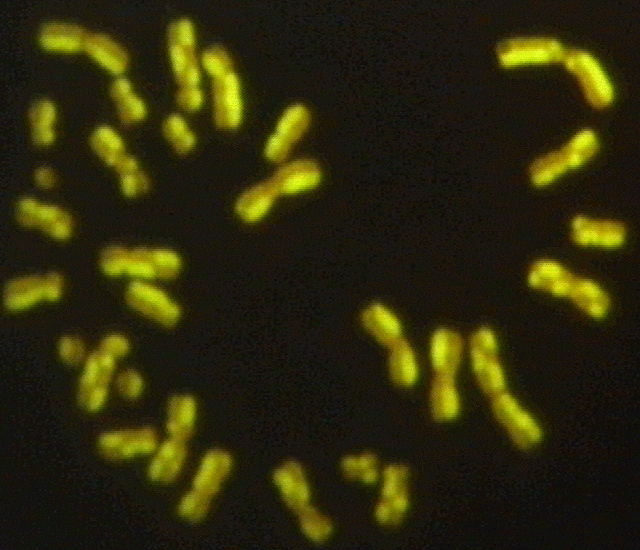
for a mutagenity test. The number of SCE is proportional to the doses of the mutagene. Below are shown mink chromosomes
carrying SCE. The SCE
chromosome is also called Harlequin chromosome. The chromosomes, without occurance
of SCE, show no breaks in the chromosome, i.e. it has two unbroken strings, one light and
one darkly stained. Where SCE occurs the chromatids change their colour. For SCE
to occur both strings of the DNA must have been broken to joint up
with the opposite string.
| Figure 10.6. Mink chromosomes with 30 XY. Showing sister chromatin exchange (SCE). |
 |